GRADE I:
- Insufficient sebaceous secretion and / or dehydration of the horny layer.
- It is especially common in women.
- Causes: inherent to the person or by external factors, which alter the skin barrier.
- Integrity of the horny layer.
the skin
the main barrier
The skin constitutes the largest organ in the human body, with a surface area of nearly 2m² and a weight that represents approximately 6% of total body weight.
The skin separates the external environment from the body’s internal environment, but this separation is not isolating, because interchanges take place between external agents and the bodily functions that contribute to shaping the appearance of the skin.

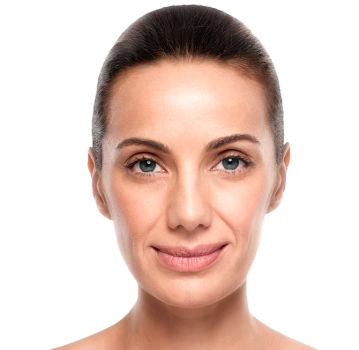
Aging represents a set of transformations that take place as a consequence of the time lived and how it has been lived. Skin aging is a reflection of the biological age, which does not always correspond with the chronological age and becomes apparent over the years through the visible signs that are: wrinkles, flaccidity or spots.
Depending on the combination of genetic factors and external factors, the process of aging will be more or less noticeable.
The Glogau scale to classify the degree of aging, takes into account aspects such as the chronological age and the influence of photoaging, to determine at what stage of aging we are and to establish the appropriate guidelines for care.
GRADE I:

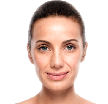
Grade II:
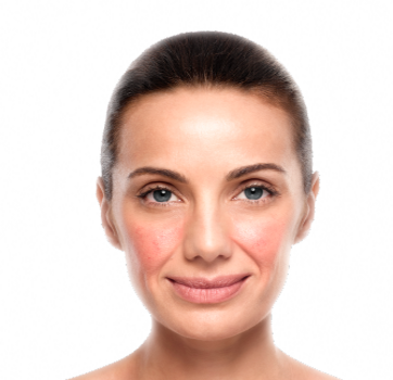
Grade III:

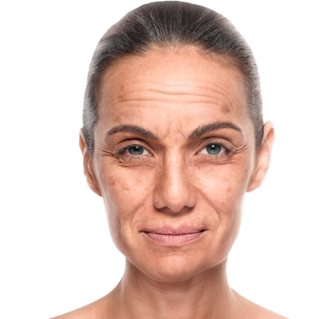
Grade IV:
Melasma:
Melasma or cloasma is an acquired hypermelanosis that manifests in areas exposed to the sun, particularly the face. They are dark brown spots, extensive, irregular in shape that appear on the face, forehead and upper lip. They appear due to hormonal changes that stimulate the melanocytes.
The usually appear in pregnant women, users of OCPs and menopausal women. This alteration is made worse by the sun.
There are two types:

Solar Lentigines:
Solar lentigines are spots on the skin associated with aging and exposure to ultraviolet radiation from the sun. They vary in color, from light brown to red or even black, and are located in the areas most exposed to the sun, particularly the hands, face, shoulders, arms and forehead, even the head if it is hairless.
From the age of 40, the skin starts to lose its ability to regenerate and recover from exposure to the sun, and the solar lentigines are very common in this age range, especially in those who spend time exposed to the sun’s rays.
Senile Lentigo:
Senile lentigines are brown to dark brown colored macules, from a few millimeters to 1-2 cm in diameter. Their surface is smooth and well defined. They usually appear after the age of 40 due to the cumulative effect of the sun and because with age melanin is unevenly distributed in the epidermis.
They are more frequent in the areas exposed to the sun like the face, back of the hands and neckline. They appear as flat, brown colored spots, and are predominantly oval in shape.

Ephelides:
Ephelides are a congenital alteration of the pigmentation that are revealed by exposure to the sun. They are commonly called freckles.
They are macules of a few millimeters diameter, light yellow or light brown, which usually appear in people with red hair or blondes with light-colored eyes. Their number increases with age.
They are located mainly on the face, neck, forearms and legs, covering the shoulders, arms and thighs during the summertime.

Microcirculation is the circuit used by the organism to transport nutrients to the tissues and to eliminate cell debris and waste substances.
Fragility, a tendency to redden and suffer irritations and above all the possibility of developing hyperactivity are characteristics of sensitive skins. They are due to alterations in the barrier function, which produces a special sensitivity of the epidermis to stimuli that can be classified into two groups:
- External stimuli: the climate, light, cosmetics, pollution, etc.
- Internal stimuli, such as stress, individual conditions, tiredness, etc.
Erythema:
Erythema is a reddening of the skin due to an excess blood supply produced by vasodilation; it is a symptom of different skin conditions; it is usually the most visible sign of a skin process that determines its size.
We may come across two types of erythema:
Solar erythema (induced), with the symptoms:
Blushing (spontaneous), with the symptoms:

Erythrosis:
This is reddening of the face (mainly the midface area). Its origin is a slowdown of the venous circulation.
The triggers can be hot drinks or food, the changes in temperature or emotions. At first it is transient, but with time the redness becomes permanent.
Couperosis:
Couperosis is an alteration of the vascularization of the dermis of the face and neckline which is expressed in the onset of redness. It corresponds to a network of telangiectasia on a background of erythrosis.
It is more common in skins that are thin, white, sensitive and pallid, which redden easily because their epidermis is very thin. The elasticity of these peripheral vessels is virtually zero. If the blood flow suddenly increases and the capacity for elastic recovery is scarce, this redness may easily occur.
The formation of couperosis is influenced by external factors (chemical agents, contusions, environmental exposure) and internal ones (digestive disorders, nervousness, stress,...).

Rosacea:
Rosacea is a vascular disease of the face that appears as a consequence of a long evolution in four stages:
This condition can be confused with and in some cases coexist with acne vulgaris and/or seborrheic dermatitis.
Rosacea affects both sexes, but is almost three times more common in women (high incidence during the menopause) and has a developmental age between 20 and 60 years of age.
The presence of reddening in the scalp or ears suggests a different diagnosis or other concomitant conditions, since rosacea is predominantly facial.

Acne is a condition with a very high incidence. Between 80% and 90% of the population suffer from it during their lifetime, the incidence among people between ages 12 to 18 is 74%.
It is caused by an excess of androgen and is the result of different trigger factors. It is characterized by a polymorphic skin condition with various types of lesion. The main factors are: excess of sebaceous secretion, reaction to the normal bacteria found on the skin, obstruction of the pilosebaceous unit.
According to the degree of development, it can have psychological and social implications, that worsen the quality of life.
Mild:
Comedones and papules

Moderate:
Comedones, papules and some pustules.

Severe:
Comedones, papules, pustules and some nodules.

Very severe:
Comedones, papules, pustules, nodules and scars.

Physiological characteristics:
Visual inspection:
Tactile inspection:
Properties:
Physiological characteristics:
Visual inspection:
Tactile inspection:
Properties:
Physiological characteristics:
Visual inspection:
Tactile inspection:
Properties: